Monday, April 25, 2011

Objective 79: Structures of the reproductive systems

Locate the structures of the male and female reproductive systems.....

http://www.labscientificequipments.com/human-reproductive-system-654.html

This was really a pretty self explanatory objective!

Objective 75: The Menstrual Cycle

Explain what happens duing follicular, ovulatory, and luteal phases of the menstrual cycle...


Ok, I embarrassed to admit this, but I had no idea.  No wonder we're so cranky when our periods come around...all that extra work!  Seriously though, I understood that during menstruation women shed their uterine lining and that was what caused their period.  I was also aware of ovulation and that it occurred typically ten days to two weeks after the period stopped and that was when a woman was most likely to get pregnant.  However, I really couldn't have given you a step by step of the process until now. 

Nurse's Note:  Oral contraceptives also known as "The Pill" prevent pregnancy when taken on a regular schedule by preventing the ovulation phase of the menstrual cycle.  Without ovulation there is no egg to be fertilized.  Other common methods of birth control include patches, rings, IUD's or intra-uterine devices.  All essentially work under the same premise of preventing ovulation and ultimately each individual should choose the method that they are most comfortable with.

Objective 73: Oogenesis and Spermatogenesis

Compare and Contrast Oogenesis and Spermatogenesis......
Spermatogenesis and Oogenesis
Meiosis, the process by which gametes are formed, can also be called gametogenesis, literally “creation of gametes.” The specific type of meiosis that forms sperm is called spermatogenesis, while the formation of egg cells, or ova, is called oogenesis. The most important thing you need to remember about both processes is that they occur through meiosis, but there are a few specific distinctions between them.
Spermatogenesis
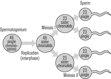
The male testes have tiny tubules containing diploid cells called spermatogonium that mature to become sperm. The basic function of spermatogenesis is to turn each one of the diploid spermatogonium into four haploid sperm cells. This quadrupling is accomplished through the meiotic cell division detailed in the last section. During interphase before meiosis I, the spermatogonium’s 46 single chromosomes are replicated to form 46 pairs of sister chromatids, which then exchange genetic material through synapsis before the first meiotic division. In meiosis II, the two daughter cells go through a second division to yield four cells containing a unique set of 23 single chromosomes that ultimately mature into four sperm cells. Starting at puberty, a male will produce literally millions of sperm every single day for the rest of his life.
Oogenesis
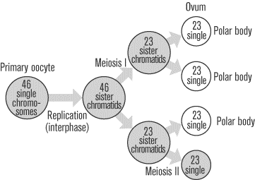
Just like spermatogenesis, oogenesis involves the formation of haploid cells from an original diploid cell, called a primary oocyte, through meiosis. The female ovaries contain the primary oocytes. There are two major differences between the male and female production of gametes. First of all, oogenesis only leads to the production of one final ovum, or egg cell, from each primary oocyte (in contrast to the four sperm that are generated from every spermatogonium). Of the four daughter cells that are produced when the primary oocyte divides meiotically, three come out much smaller than the fourth. These smaller cells, called polar bodies, eventually disintegrate, leaving only the larger ovum as the final product of oogenesis. The production of one egg cell via oogenesis normally occurs only once a month, from puberty to menopause.


So I was reading through the textbook, trying to get a good handle on the processes of oogenesis and spermatogenesis.  Theres alot of information involved in these two processes, so I went online to see what  else I could find whether it be a comparative chart or a video etc. I ended up putting this chart together after doing quite a few reviews of the information.  After the time spent reading and the time spent online I basically came to one conclusion....  NATURALLY IT TAKES FOUR MEN (OR MALE PARTS) TO DO WHAT ONE WOMAN CAN DO!!!!!!


Oogenesis and Spermatogenesis – Comparative Chart

OOGENESIS


SPERMATOGENESIS
Oogonium
Female germ cell

Spermatogonium
Male germ cell

Germ cells committed to Meiosis
Primary Oocyte


Primary Spermatocyte
↙↘

First Meiotic Division
Secondary                First
Oocyte                  Polar Body

Secondary             Secondary
Spermatocyte    Spermatocyte

Second Meiotic Division
                            ↙↘
Ovum and Second Polar Body


4 Spermatids


Spermiogenesis
1 Ovum
1 viable gamete

4 Spermatazoa
4 viable gametes

Sunday, April 24, 2011

Objectives 71 & 72: How spermatazoa move and evolutionary fitness

Exploring how spermatazoa move through the female reproductive tract and evaluating fertilization in terms of evolutionary fitness......


Well, I guess in a way, sperm are pretty incredible.  When you look at the resistance that they have to overcome in order to achieve their "goal" and the way that they're structured in order to make that happen ...overall, pretty amazing.  Lets take a head to toe look at the structure of the sperm.  At the head, we find the nucleus which encloses the most densely packed dna found in any of the eukaryote cells.  More interesting is that the head is enclosed in an acrosome that contains enzymes that will help it to break down the egg when or if it ever reaches one.  Another interesting fact regarding this is that the acrosome is actually alkaline in nature, so the acidity of the vagina is yet another hurdle that it must overcome.  However, it does have glycoproteins on that acrosome that all immune systems recognize allowing it to enter and find its way through the body without being attacked.


The term spermatzoan actually only applies to a sperm that is mobile.  A large part of that mobility stems from the midpiece of the sperm just below the head that is actually surrounded by ATP to provide energy for movement.  And finally the tail, which I kind of relate to one of those super small 15 horsepower motors on a john-boat, it works and it spins, but it sure itsn't gonna get you anywhere fast!

So it really is amazing that the sperm even makes it to the egg in order to fertilize it.  The fact that it does relates to evolutionary fitness to me.  It will be the strongest, the fastest, the "smartest" sperm that reaches the egg.  The rest simply die.  In my mind this is reflective of evolution itself.  Only the strong survive.

Objectives 74, 76 & 77: Zygotes, Morula and Blastocyst

Distinguishing between diploid germ cells and haploid sex cells, differentiating between a zygote, morula, and blastocyst and recognizing that a blastocyst secretes human gonadotropin, which prolongs the life of the corpus luteum....


Well, I kind of lucked out looking for this video while I was actually looking for a video of some swimming sperm!  (Lucky me)!  Anyway, the video contained the basic information covering objectives 76 and 77, but I think that objective 74 can be fit nicely in here as well, so here goes!  The video gave a step by step starting from the time that the zygote had actually implanted itself in the endometrium lining of the uterus wall.  HERES THE THING:  What it did not mention is what a zygote is.  A zygote is actually two haploid sex cells fused together to form a diploid cell that will hopefully someday become a baby.  This goes back to Bio-100 again, where we studied our genetic makeup. So this is how that works, it actually takes a full 46 chromosomes to make a human being.  When the gametes or sex cells are produced during meiosis, they are produced with only 23 chromosomes.  This way when the two gametes (the sperm and the ova or egg) join together, the future fetus that they produce will have genetic characteristics from both parents. 

So in brief, a haploid cell has 23 chromosomes (think Haploid = Half), where as a diploid cell has a full 46 chromosomes (think Diploid = Double)!

To talk just a little bit more about this, I again refer back to the iconic Bio-100!  I specifically remember Mrs. Gess using her hands to form a ball (a morula, the ball of divided cells)) and then beginning to move her hands inward to form the blastocyst....a tube within a tube.

Even more amazing after learning this information is that that zygote which then becomes the morula and then becomes a blastocyst then becomes:

This fetus ......
who then becomes....this baby...

who then becomes....this little girl.
~ Aubrey Grayson Kersey ~

When you look at it from that perspective, its awe inducing~

Saturday, April 23, 2011

Objective 78: The Alimentary Canal

Distinguish among the different parts (gross and histological) of the alimentary canal in terms of structure and function.................
This picture from the Anatomy and Physiology textbook* explains the basic layers of all portions of the alimentary canal.  One of the greatest hurdles to overcome with the alimentary canal is the understanding that while all layers have an internal epithelium for the mucosa, each of those layers will have a different type of epithelium that relates to the structures function.  For example the mouth and esophagus have an epithelium layer of stratified squamous to protect them from abrasion.  The stomach and the small intestine are both lined with simple columnar epithelium yet the stomach epithelium is designed for secretion of gastric juices while the small intestines epithelium is designed for absorption.

Nurse's Note: Damage to the stomach's epithelial lining can lead to gastric ulcers which can cause severe stomach pain.  Once thought to be caused by certain medications and ingesting spicey food, the H.Pylori bacteria is now thought to be the most common cause.

Objective 57: Lacteals

Justify the role of lacteals in transporting products of digestion..............

A lacteal is a lymphatic capillary that absorbs dietary fats in the villi of the small intestine. http://en.wikipedia.org/wiki/Lacteal


http://en.wikipedia.org/wiki/Lacteal

I'm really not sure how to reflect on this except to say that it seems pretty basic.  The villi in the small intestine contain both a lacteal or lymph capillary and a capilary bed.  The digested food is absorbed into the epithelium and sent throughout the body via the lymph capillary and the capillary bed. 

Also, this lacteal picture reminds me of a sunflower !